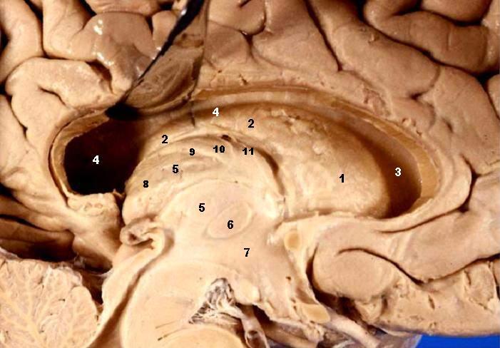
{{Information| |Description='''Human brain left dissected - midsagittal view''' Fornix & Septum Pellucidum Resected – Lateral Ventricle Exposed The head and body of the Caudate nucleus produce a large elevation in the lateral wall of the anterior hor
ไฟล, human, brain, left, dissected, midsagittal, view, description, ไฟล, ประว, ไฟล, หน, าท, ภาพน, การใช, ไฟล, วนกลางไม, ภาพท, รายละเอ, ยดส, งกว, าน, human, brain, left, dissected, midsagittal, view, description, 8206, กเซล, ขนาดไฟล, โลไบต, ชน, ดไมม, image, jpe. ifl prawtiifl hnathimiphaphni karichiflswnklangimmiphaphthimiraylaexiydsungkwani Human brain left dissected midsagittal view description 2 JPG 8206 701 488 phikesl khnadifl 50 kiolibt chnidimm image jpeg rupphaphhruxiflesiyngni tnchbbxyuthi khxmmxns raylaexiyddanlang epnkhxkhwamthiaesdngphlcak ifltnchbbinkhxmmxns khxmmxnsepnewbistinokhrngkarsahrbekbrwbrwmsuxesri thi khunsamarthchwyid khwamyx khaxthibayHuman brain left dissected midsagittal view description 2 JPG Human brain left dissected midsagittal viewFornix amp Septum Pellucidum Resected Lateral Ventricle ExposedThe head and body of the Caudate nucleus produce a large elevation in the lateral wall of the anterior horn and body of the lateral ventricle Nucleus caudatus Caput Nucleus caudatus Corpus Ventriculus lateralis Cornu frontale Ventriculus lateralis Pars centralis Thalamus Adhaesio interthalamica Hypothalamus Ventriculus tertius Pulvinar thalami Taenia choroidea Lamina affixa Stria terminalis font arial black size 10 wnthi 30 phvscikayn ph s 2548aehlngthima http www healcentral org healapp showMetadata metadataId 40566 Internet Archive of file description page phusrangsrrkh John A Beal PhD Dep t of Cellular Biology amp Anatomy Louisiana State University Health Sciences Center Shreveportkarxnuyat karichiflniihm CC BYewxrchnxun Human brain left dissected midsagittal view JPG karxnuyatichsiththi iflnixyuphayitsyyaxnuyatkhriexthifkhxmmxns runaesdngthima 2 5 thwipkhunsamarth thicaaebngpn thicathasaena aeckcay aelasngngandngklawtxip thicaeriyberiyngihm thicaddaeplngngandngklaw phayitenguxnikhtxipni aesdngthima khuntxngihekiyrtiecakhxngnganxyangehmaasm odyephimlingkipyngsyyaxnuyat aelarabuhakmikarepliynaeplng khunxacthaechnniidinrupaebbidkidtamkhwr aettxngimichinlksnathiaenawaphuihxnuyatsnbsnunkhunhruxkarichngankhxngkhunhttps creativecommons org licenses by 2 5 CC BY 2 5 Creative Commons Attribution 2 5 true true This file which was originally posted to http www healcentral org healapp showMetadata metadataId 40566 was reviewed on 25 September 2013 by reviewer Eleassar who confirmed that it was available there under the stated license on that date AnnotationsInfoFieldThis image is annotated View the annotations at Commons460 231 37 39 701 488 Nucleus caudatus Caput 216 176 29 39 701 488 Nucleus caudatus Corpus 536 222 42 37 701 488 Ventriculus lateralis Cornu frontale 116 201 38 46 701 488 Ventriculus lateralis Pars centralis 311 301 23 27 701 488 Adhaesio interthalamica 337 343 19 23 701 488 Hypothalamus Ventriculus tertius 192 245 24 25 701 488 Pulvinar thalami 269 278 26 28 701 488 Thalamus 258 202 22 23 701 488 Taenia choroidea 293 194 28 30 701 488 Lamina affixa 331 197 35 34 701 488 Stria terminaliskhabrryayodyyxithyephimkhabrryaythrrthdediywephuxkhyaykhwamwaiflnimixairixethmthiaesdngxyuiniflniprakxbdwysthanalikhsiththimilikhsiththisyyaxnuyatCreative Commons Attribution 2 5 Generic xngkvswnthisrang wnkxtng30 phvscikayn 2005 prawtiifl khlikwnthi ewlaephuxduiflthipraktinkhnann wnthi ewlarupyxkhnadphuichkhwamehn pccubn05 14 23 mithunayn 2549701 488 50 kiolibt Patho Information Description 039 039 039 Human brain left dissected midsagittal view 039 039 039 Fornix amp Septum Pellucidum Resected Lateral Ventricle Exposed The head and body of the Caudate nucleus produce a large elevation in the lateral wall of the anterior hor hnathimiphaphni hnatxipni oyngmathiphaphni klumniwekhliys pulvinar niwekhliysmihang karichiflswnklang wikixuntxipniichiflni karichbn ar wikipedia org الغدة النخامية وعلاقتها بالوطاء karichbn bg wikipedia org Hipotalamus karichbn de wikibooks org Neuroanatomie Zwischenhirn Neuroanatomie Druckversion karichbn en wikipedia org Hypothalamus Pulvinar nuclei Lamina affixa karichbn es wikipedia org Estria terminal karichbn fa wikipedia org هیپوتالاموس بطن های طرفی karichbn fr wikipedia org Noyau caude karichbn ja wikipedia org 側脳室 尾状核 karichbn pa wikipedia org ਹ ਪ ਥ ਲ ਮਸ karichbn ro wikipedia org Hipotalamus karichbn ru wikipedia org Podushka talamusa Perednee yadro podushki talamusa Nizhnee yadro podushki talamusa Lateralnoe yadro podushki talamusa Medialnoe yadro podushki talamusa karichbn sh wikipedia org Hipotalamus karichbn sl wikipedia org Cloveski mozgani ekhathungcak https th wikipedia org wiki ifl Human brain left dissected midsagittal view description 2 JPG, wikipedia, วิกิ หนังสือ, หนังสือ, ห้องสมุด,