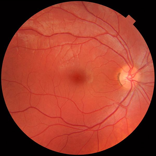
English: Fundus photograph of the right eye, showing a fundus with no sign of disease or pathology. It is seen from front so that left in each image is to the person's right. The gaze is into the camera, so the macula is in the center of the image, and the optic disk is located towards the nose (right in image). The optic disk has some pigmentation at the perimeter of the lateral side, which is considered non-pathological.
Veins are darker and slightly wider than corresponding arteries. Major nerve pathways are seen as white striped patterns radiating from the optic disk. In addition, there are also lighter areas close to larger vessels seen mainly at upper left in the image (person's upper right), which is regarded as a normal finding in younger people.
Photo is taken at Gävle Hospital in Sweden in 2012 on a healthy 25-year old male volunteer.
วันที่
แหล่งที่มา
งานของตัว
ผู้สร้างสรรค์
Mikael Häggström.
When using this image in external works, it may be cited as:
Häggström, Mikael (2014). "Medical gallery of Mikael Häggström 2014". WikiJournal of Medicine1 (2). DOI:10.15347/wjm/2014.008. ISSN 2002-4436. Public Domain.
ไฟล์นี้มีให้ใช้ภายใต้ CC0 1.0 Universal Public Domain Dedication ของครีเอทีฟคอมมอนส์
The person who associated a work with this deed has dedicated the work to the public domain by waiving all of their rights to the work worldwide under copyright law, including all related and neighboring rights, to the extent allowed by law. You can copy, modify, distribute and perform the work, even for commercial purposes, all without asking permission.
http://creativecommons.org/publicdomain/zero/1.0/deed.enCC0Creative Commons Zero, Public Domain Dedicationfalsefalse
WikiJournal of Medicine/Medical gallery of Mikael Häggström 2014
การใช้บน fr.wikiversity.org
Introduction à l'infographie/Notions Fondamentales
視覺系統
視覺皮層
มกราคม 18, 2023
ไฟล, fundus, photograph, normal, right, ไฟล, ประว, ไฟล, หน, าท, ภาพน, การใช, ไฟล, วนกลางขนาดของต, วอย, างน, กเซล, ความละเอ, ยดอ, กเซล, กเซล, กเซล, กเซล, กเซล, ภาพท, ความละเอ, ยดส, งกว, 8206, กเซล, ขนาดไฟล, โลไบต, ชน, ดไมม, image, jpeg, ปภาพหร, อไฟล, เส, ยงน, น. ifl prawtiifl hnathimiphaphni karichiflswnklangkhnadkhxngtwxyangni 600 600 phikesl khwamlaexiydxun 240 240 phikesl 480 480 phikesl 768 768 phikesl 1 024 1 024 phikesl 1 411 1 411 phikesl duphaphthimikhwamlaexiydsungkwa 8206 1 411 1 411 phikesl khnadifl 248 kiolibt chnidimm image jpeg rupphaphhruxiflesiyngni tnchbbxyuthi khxmmxns raylaexiyddanlang epnkhxkhwamthiaesdngphlcak ifltnchbbinkhxmmxns khxmmxnsepnewbistinokhrngkarsahrbekbrwbrwmsuxesri thi khunsamarthchwyid khwamyx khaxthibayFundus photograph of normal right eye jpg English Fundus photograph of the right eye showing a fundus with no sign of disease or pathology It is seen from front so that left in each image is to the person s right The gaze is into the camera so the macula is in the center of the image and the optic disk is located towards the nose right in image The optic disk has some pigmentation at the perimeter of the lateral side which is considered non pathological Veins are darker and slightly wider than corresponding arteries Major nerve pathways are seen as white striped patterns radiating from the optic disk In addition there are also lighter areas close to larger vessels seen mainly at upper left in the image person s upper right which is regarded as a normal finding in younger people Photo is taken at Gavle Hospital in Sweden in 2012 on a healthy 25 year old male volunteer wnthi 21 minakhm ph s 2555aehlngthima ngankhxngtwphusrangsrrkh Mikael Haggstrom When using this image in external works it may be cited as Haggstrom Mikael 2014 Medical gallery of Mikael Haggstrom 2014 WikiJournal of Medicine 1 2 DOI 10 15347 wjm 2014 008 ISSN 2002 4436 Public Domain or By Mikael Haggstrom used with permission ewxrchnxun Left eye karxnuyatichsiththi khapheca inthanaphuthuxlikhsiththikhxngphaphhruxsuxni xnuyatihichphaphhruxsuxniphayitenguxnikhtxipni iflnimiihichphayit CC0 1 0 Universal Public Domain Dedication khxngkhriexthifkhxmmxnsThe person who associated a work with this deed has dedicated the work to the public domain by waiving all of their rights to the work worldwide under copyright law including all related and neighboring rights to the extent allowed by law You can copy modify distribute and perform the work even for commercial purposes all without asking permission http creativecommons org publicdomain zero 1 0 deed en CC0 Creative Commons Zero Public Domain Dedication false falsekhabrryayodyyxithyephimkhabrryaythrrthdediywephuxkhyaykhwamwaiflnimixairxngkvsFundus photograph of the right eye showing a fundus with no sign of disease or pathology ixethmthiaesdngxyuiniflniprakxbdwydwngtacxtafundus xngkvscksuwithyasthanalikhsiththicopyrighted dedicated to the public domain by copyright holder xngkvssyyaxnuyatCC0 xngkvswnthisrang wnkxtng21 minakhm 2012thimakhxngiflkarsrangdngedimodyphuxpohld prawtiifl khlikwnthi ewlaephuxduiflthipraktinkhnann wnthi ewlarupyxkhnadphuichkhwamehn pccubn17 49 21 minakhm 25551 411 1 411 248 kiolibt Mikael Haggstrom hnathimiphaphni hnatxipni oyngmathiphaphni karthayphaphcxprasathta tamnusy karichiflswnklang wikixuntxipniichiflni karichbn ar wikipedia org شبكية قاع العين karichbn bs wikipedia org Ocno dno karichbn ca wikipedia org Fons de l ull Retinografia karichbn de wikipedia org Morbus Coats karichbn en wikipedia org Retina Macula Human eye Ophthalmoscopy Fundus eye Fundus photography User Mikael Haggstrom Gallery karichbn en wikiversity org WikiJournal of Medicine Medical gallery of Mikael Haggstrom 2014 karichbn et wikipedia org Karotenoidid karichbn fr wikipedia org Fond d œil karichbn fr wikiversity org Introduction a l infographie Notions Fondamentales karichbn gl wikipedia org Macula lutea karichbn he wikipedia org מחלת קואוטס karichbn hu wikipedia org Erhartya karichbn it wikipedia org Fundus karichbn ml wikipedia org റ റ റ ന karichbn pl wikipedia org Leukokoria karichbn uk wikipedia org Zorovij nerv karichbn zh yue wikipedia org 視覺 視覺系統 視覺皮層 karichbn zh wikipedia org 眼底 ekhathungcak https th wikipedia org wiki ifl Fundus photograph of normal right eye jpg, wikipedia, วิกิ หนังสือ, หนังสือ, ห้องสมุด,